Current issue
Archive
Online first
About the Journal
Editorial Board
Scientific Board
Reviewers
Abstracting and indexing
Publisher
Contact
Subscription
Ethical standards and procedures
Abstracting and indexing
For authors
Ethical standards and procedures
Publication charge
Books and Events
Books
Events
CASE REPORT
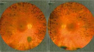
Macular Hole of the Left Eye in a 41-year-old Patient with Retinitis pigmentosa. A Case Report
1
Students’ Scientific Society, Department of Ophthalmology, Faculty of Medical Sciences in Katowice, Medical University of Silesia in Katowice, Poland
2
Department of Ophthalmology, Faculty of Medical Sciences in Katowice, Medical University of Silesia in Katowice, Poland
Head: Professor Ewa Mrukwa-Kominek, MD, PhD
3
Kornel Gibiński University Clinical Centre in Katowice, Poland
Head: Professor Ewa Mrukwa-Kominek, MD, PhD
Submission date: 2024-01-08
Acceptance date: 2024-02-19
Publication date: 2024-05-02
Ophthalmology 2024;27(1):24-26

KEYWORDS
ABSTRACT
Retinitis pigmentosa is characterized by degeneration of the photoreceptors or retinal pigment epithelium and causes progressive vision loss. The disease can lead to night blindness, reduced field of vision and finally to complete loss of vision. The report describes a case of a patient diagnosed with retinitis pigmentosa who was admitted to hospital for further diagnosis and treatment. For several months, the patient reported a gradual decrease in visual acuity, especially in the left eye and visual impairments in poor lighting. Retinitis pigmentosa is a genetic disorder, therefore genetic counseling and screening of family members for retinitis pigmentosa is important. The specific pharmacological treatment is still unknown. In severe cases posterior vitrectomy is performed as a basic method of curing macular holes.
REFERENCES (16)
2.
Ferrari S, Di Iorio E, Barbaro V, et al.: Retinitis pigmentosa: genes and disease mechanisms. Curr Genomics. 2011 Jun; 12(4): 238–249.
3.
Berson EL, Sandberg MA, Rosner B, et al.: Natural course of retinitis pigmentosa over a three-year interval. American Journal of Ophthalmology. 1985; 99(3): 240–251.
4.
Flynn MF, Fishman GA, Anderson RJ, et al.: Retrospective longitudinal study of visual acuity change in patients with retinitis pigmentosa. Retina 2001 Dec; 21(6): 639–646.
5.
Fahim A: Retinitis pigmentosa: recent advances and future directions in diagnosis and management. Curr Opin Pediatr. 2018 Dec; 30, 6: 725–733.
6.
Vingolo EM, Valente S, Gerace E, et al.: Macular hole in retinitis pigmentosa patients: microincision vitrectomy with polydimethylsiloxane as possible treatment. Eye (Lond). 2015 May 29; 5: 699–702.
7.
Aizawa S, Mitamura Y, Baba T, et al.: Correlation between visual function and photoreceptor inner/outer segment junction in patients with retinitis pigmentosa. Eye (Lond). 2009 Feb; 23(2): 304–308. doi: 10.1038/sj.eye.6703076. Epub 2008 Jan 11. PMID: 18188175.
8.
Wakabayashi T, Oshima Y, Fujimoto H, et al.: Foveal microstructure and visual acuity after retinal detachment repair: imaging analysis by Fourier-domain optical coherence tomography. Ophthalmology. 2009 Mar; 116(3): 519–528. doi: 10.1016/j.ophtha.2008.10.001. Epub 2009 Jan 14. PMID: 19147231.
9.
Sahel JA, Marazova K, Audo I: Clinical characteristics and current therapies for inherited retinal degenerations. Cold Spring Harb Perspect Med. 2014 Oct 16; 5(2): a017111. doi: 10.1101/cshperspect.a017111. PMID: 25324231; PMCID: PMC4315917.
10.
Cross N, van Steen C, Zegaoui Y, et al.: Current and Future Treatment of Retinitis Pigmentosa. Clin Ophthalmol. 2022 Aug 31; 16: 2909–2921. (7).
11.
Gagliardi G, M’Barek KB, Goureau O: Photoreceptor cell replacement in macular degeneration and retinitis pigmentosa: a pluripotent stem cell-based approach. Prog Retin Eye Res. 2019 Jul; 71: 1–25. (8).
12.
Huang XF: Current Pharmacological Concepts in the Treatment of the Retinitis Pigmentosa. Adv Exp Med Biol. 2018 May; 1074: 439–445.
13.
European Physician Research. Lightning health European ophthalmologist research; 2021.
14.
Gass JD: Idiopathic senile macular hole. Its early stages and pathogenesis. Archives of Ophthalmology. 1988 May 106; 5: 629–639.
15.
Wakely L, Rahman R, Stephenson J: A comparison of several methods of macular hole measurement using optical coherence tomography, and their value in predicting anatomical and visual outcomes. British Journal of Ophthalmology. 2012 Jul 96; 7: 1003–1007.
16.
Bikbova G, Oshitari T, Baba T, et al.: Pathogenesis and Management of Macular Hole: Review of Current Advances. J Ophthalmol. 2019 May 2; 2019: 3467381.
Share
RELATED ARTICLE
| eISSN: | 1689-362X |
| ISSN: | 1505-2753 |
We process personal data collected when visiting the website. The function of obtaining information about users and their behavior is carried out by voluntarily entered information in forms and saving cookies in end devices. Data, including cookies, are used to provide services, improve the user experience and to analyze the traffic in accordance with the Privacy policy. Data are also collected and processed by Google Analytics tool (more).
You can change cookies settings in your browser. Restricted use of cookies in the browser configuration may affect some functionalities of the website.
You can change cookies settings in your browser. Restricted use of cookies in the browser configuration may affect some functionalities of the website.