OPIS PRZYPADKU
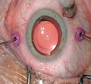
Minimally Invasive Vitreoretinal Surgery (25G, 27G) with High-speed Vitrectomy Cutter – Case Series
1
Department of Ophthalmology Medical University of Gdańsk
Head: Professor Katarzyna Michalska-Małecka, MD, PhD
Data nadesłania: 15-03-2024
Data akceptacji: 09-04-2024
Data publikacji: 02-05-2024
Ophthalmology 2024;27(1):29-32
SŁOWA KLUCZOWE
STRESZCZENIE
Introduction:: The development of vitreoretinal surgery is inseparably linked to the gradual miniaturization and decreasing invasiveness of the procedure. The smallest available systems are 23G, 25G, and 27G, which are referred to as minimally invasive vitreoretinal surgery techniques. This paper presents the preoperative and postoperative results of three patients undergoing posterior vitrectomy using the 25G or 27G systems with the HIPERVIT probe (Alcon, USA). The effects were retrospectively analyzed in terms of changes in best corrected visual acuity, intraocular pressure, occurrence of complications, and changes in optical coherence tomography images of macular edema. Examinations were performed before and one day, two weeks, three months, and six months after vitrectomy. The potential benefits and drawbacks of using these systems in practice were analyzed. Case report:: The analysis concerns the results of the performed posterior vitrectomy procedure in patients with full-thickness macular hole, tractional retinal detachment due to diabetic retinopathy, and vitreomacular traction syndrome. Conclusions:: The performed procedures allowed for improvement in both anatomical and functional conditions of the macula. All analyzed patients showed an improvement in visual acuity. None of the analyzed patients required the use of scleral sutures. Serious complications were not observed during the surgery or during the 6-month observation period. 25G and 27G HYPERVIT vitrectomy can be successfully used in the discussed vitreoretinal diseases. In the discussed cases, it enabled the effective and safe performance of the procedure.
REFERENCJE (20)
1.
De Juan E. Jr, Hickingbotham D: Refinements in microinstrumentation for vitreous surgery. Am J Ophthalmol. 1990; 109(2): 218–220.
2.
Fujii GY, De Juan E Jr., Humayun MS, et al.: A new 25-gauge instrument system for transconjunctivalsutureless vitrectomy system for vitreoretinal surgery. Ophtahlmology. 2002; 109(10): 1807–1812; discussion 1813.
3.
Eckardt C: Transconjunctival sutureless 23-gauge vitrectomy. Retina. 2005; 25: 208–211.
4.
Osawa S, Oshima Y: Innovations in 27-gauge vitrectomy for sutureless microincision vitrectomy surgery. Retina Today. 2014; July/ August: 42–45.
5.
Thompson JT: Advantages and limitations of small gauge vitrectomy. Surv Ophthalmol. 2011 Mar-Apr; 56(2): 162–172. doi: 10.1016/j.survophthal.2010.08.003. Epub 2011 Jan 14. PMID: 21236459.
6.
Hubschman JP, Gupta A, Bourla DH, et al.: 20-, 23-, and 25-gauge vitreous cutters: performance and characteristics evaluation. Retina. 2008 Feb; 28(2): 249–257. doi: 10.1097/IAE.0b013e31815ec2b3. PMID: 18301030.
7.
Rossi T, Querzoli G, Angelini G, et al.: Introducing new vitreous cutter blade shapes: a fluid dynamics study. Retina. 2014 Sep; 34(9): 1896–1904. doi: 10.1097/IAE.0000000000000143. PMID: 24871998.
8.
Teixeira A, Chong L, Matsuoka N, et al.: Novel method to quantify traction in a vitrectomy procedure. Br J Ophthalmol. 2010 Sep; 94(9): 1226–1229. doi: 10.1136/bjo.2009.166637. Epub 2010 Jun 10. PMID: 20538657.
9.
Jairath NK, Paulus YM, Yim A, et al.: Intra- and post-operative risk of retinal breaks during vitrectomy for macular hole and vitreomacular traction. PLoS One. 2022 Aug 11; 17(8): e0272333. doi: 10.1371/journal.pone.0272333. PMID: 35951646; PMCID: PMC9371285.
10.
Steel DH, Charles M, Zhu Y, et al.: Fluidic performance of a dual-action vitrectomy probe compared with a single-action probe. Retina. 2022 Nov 1; 42(11): 2150–2158. doi: 10.1097/IAE.0000000000003573. Epub 2022 Jul 21. PMID: 35868025; PMCID: PMC9584060.
11.
Frisina R, Pinackatt SJ, Sartore M, et al.: Cystoid macular edema after pars plana vitrectomy for idiopathic epiretinal membrane. Graefes Arch Clin Exp Ophthalmol. 2015 Jan; 253(1): 47–56. doi: 10.1007/s00417-014-2655-x. Epub 2014 May 25. PMID: 24859385.
12.
Recchia FM, Scott IU, Brown GC, et al.: Small-gauge pars plana vitrectomy: a report by the American Academy of Ophthalmology. Ophthalmology. 2010; 117(9): 1851–1857.
13.
Khan MA, Shahlaee A, Toussaint B, et al.: Outcomes of 27 gauge microincision vitrectomy surgery for posterior segment disease. Am J Ophthalmol. 2016; 161: 36–43.
14.
Oshima Y, Wakabayashi T, Sato T, et al.: A 27-gauge instrument system for transconjunctival sutureless microincision vitrectomy surgery. Ophthalmology. 2010 Jan; 117(1): 93–102.e2. doi: 10.1016/j.ophtha.2009.06.043. Epub 2009 Oct 31. PMID: 19880185.
15.
Baudin F, Benzenine E, Mariet AS, et al.: Epidemiology of Acute Endophthalmitis after Intraocular Procedures: A National Database Study. Ophthalmol Retina. 2022 Jun; 6(6): 442–449. doi: 10.1016/j.oret.2022.01.022. Epub 2022 Feb 5. PMID: 35134544.zz.
16.
Chiang A, Kaiser RS, Avery RL, et al.: Endophthalmitis in microincision vitrectomy: outcomes of gas-filled eyes. Retina. 2011 Sep; 31(8): 1513– –1517. doi: 10.1097/IAE.0b013e3182209290. PMID: 21878799.
17.
Tekin K, Sonmez K, Inanc M, et al.: Evaluation of corneal topographic changes and surgically induced astigmatism after transconjunctival 27-gauge microincision vitrectomy surgery. Int Ophthalmol. 2018; 38: 635–643.
18.
Ma J, Wang Q, Niu H: Comparison of 27-Gauge and 25-Gauge Microincision Vitrectomy Surgery for the Treatment of Vitreoretinal Disease: A Systematic Review and Meta-Analysis. J Ophthalmol. 2020 Aug 18; 2020: 6149692. doi: 10.1155/2020/6149692. PMID: 32908682; PMCID: PMC7450297.
19.
Shroff CM, Gupta C, Shroff D, et al.: Bimanual microincision vitreous surgery for severe proliferative diabetic retinopathy: outcome in more than 300 eyes. Retina. 2018 Sep; 38 Suppl 1: S134–S145. doi: 10.1097/IAE.0000000000002093. PMID: 29406470.
20.
Rossi T, Querzoli G, Angelini G, et al.: Introducing new vitreous cutter blade shapes: a fluid dynamics study. Retina. 2014 Sep; 34(9): 1896–1904.
Udostępnij
ARTYKUŁ POWIĄZANY
Przetwarzamy dane osobowe zbierane podczas odwiedzania serwisu. Realizacja funkcji pozyskiwania informacji o użytkownikach i ich zachowaniu odbywa się poprzez dobrowolnie wprowadzone w formularzach informacje oraz zapisywanie w urządzeniach końcowych plików cookies (tzw. ciasteczka). Dane, w tym pliki cookies, wykorzystywane są w celu realizacji usług, zapewnienia wygodnego korzystania ze strony oraz w celu monitorowania ruchu zgodnie z Polityką prywatności. Dane są także zbierane i przetwarzane przez narzędzie Google Analytics (więcej).
Możesz zmienić ustawienia cookies w swojej przeglądarce. Ograniczenie stosowania plików cookies w konfiguracji przeglądarki może wpłynąć na niektóre funkcjonalności dostępne na stronie.
Możesz zmienić ustawienia cookies w swojej przeglądarce. Ograniczenie stosowania plików cookies w konfiguracji przeglądarki może wpłynąć na niektóre funkcjonalności dostępne na stronie.


